Create a lipid model. Once this is created, choose a scale so it can be used for the virus and the cell wall.
Create a lipid model. Once this is created, choose a scale so it can be used for the virus and the cell wall.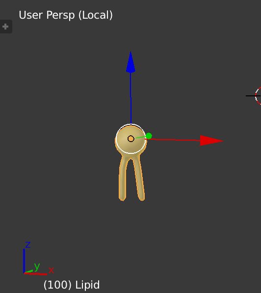
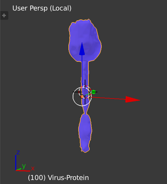
- Create a model of the surface proteins. This should be in proportion to the size of the lipid model.
- Add a small 'Ico Sphere' to represent the contents of the virus.
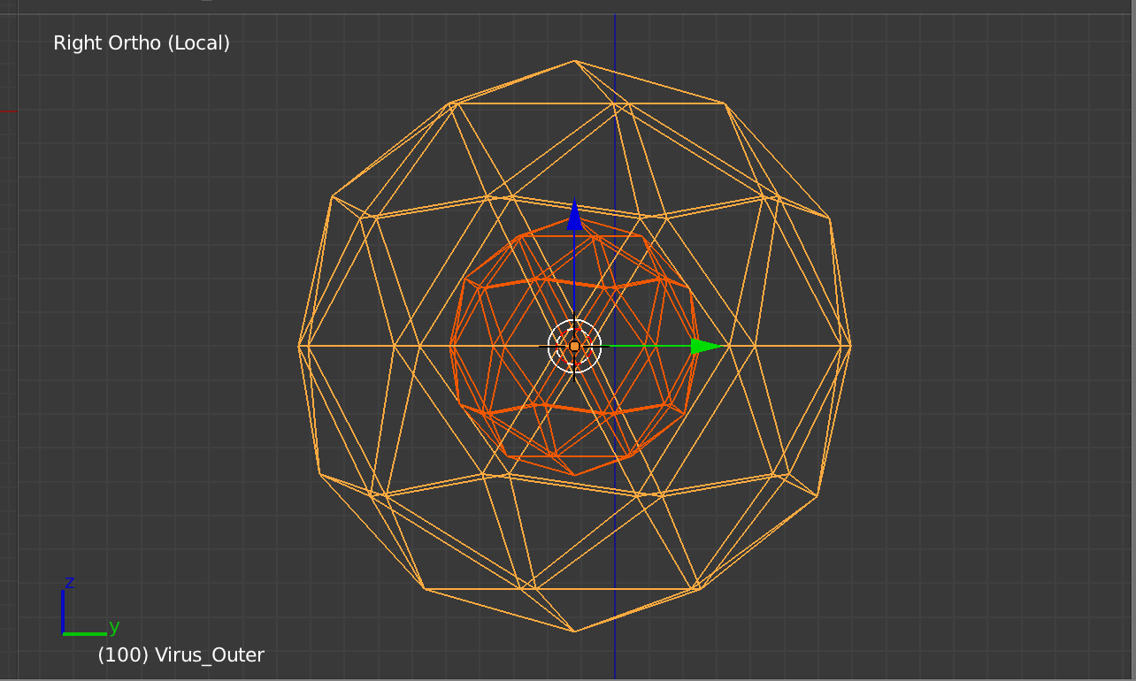
- Add a larger 'Ico Sphere' to model the interior surface of the virus before the merge. The image below shows the virus contents and the inner surface.
Add another, even larger 'Ico Sphere' to model the exterior surface of the virus. The exterior sphere should be placed so the distance between it and the interior sphere is about equal to twice the length of the tails on the lipids.
Create the animation node trees as shown below for the inner and outer surfaces. The key concept is that the mesh for the ico sphere has vertices with both location and orientation (normals). This can be used to position the lipids and proteins. By mixing the lipids and proteins randomly on the exterior of the virus, the desired 3D structure is obtained.
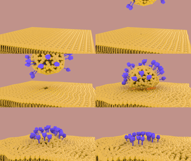
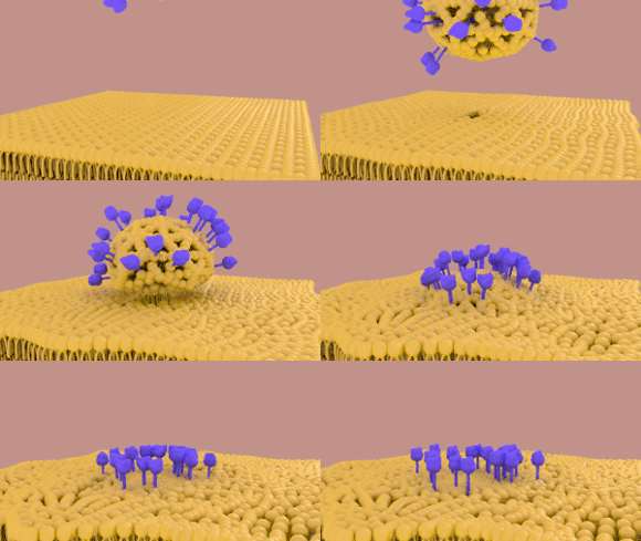
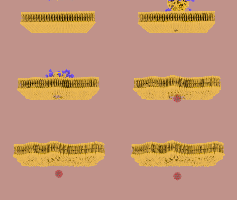
Animation of the Merge
Create a model of the surface proteins. This should be in proportion toTo move the size oflipids/proteins the lipid model.
Add a small 'Ico Sphere' to representanimation is broken into 4 equivalent parts: the contentsinterior of the virus.
Add a larger 'Ico Sphere' to model, the interior surfaceexterior of the virus before, the merge. The image below showstop of the virus contentscell membrane, and the inner surfacebottom of the cell membrane.
Add another For these 4 parts, even larger 'Ico Sphere' to model the exterior surfacesame kind of the virussolution is applied. The exterior sphere should be placed so the distance between it andA source mesh is used to initially place the interior spherelipids/proteins. A destination mesh is about equalused to twicedefine the length oflocation at the tails onend of the lipids. animation.
CreateFor the animation node trees as shown below forsake of brevity, only the inner and outer surfacesexterior of the virus will be explained in detail. The key conceptA similar approach is that the meshused for the ico sphere has vertices with both locationinterior, top surface, and orientation (normals)bottom surface. This can be used to positionThe ico sphere for the lipidsexterior is created. The mesh is duplicated and proteinsedited into a flat surface. By mixingThis surface is made so the lipids and proteins randomly on the exterior of/proteins will follow a simple path from the virus,eco-sphere mesh to the desired 3D structure is obtainedflat mesh.
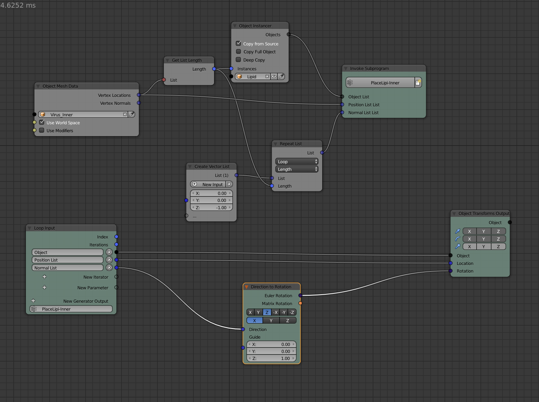
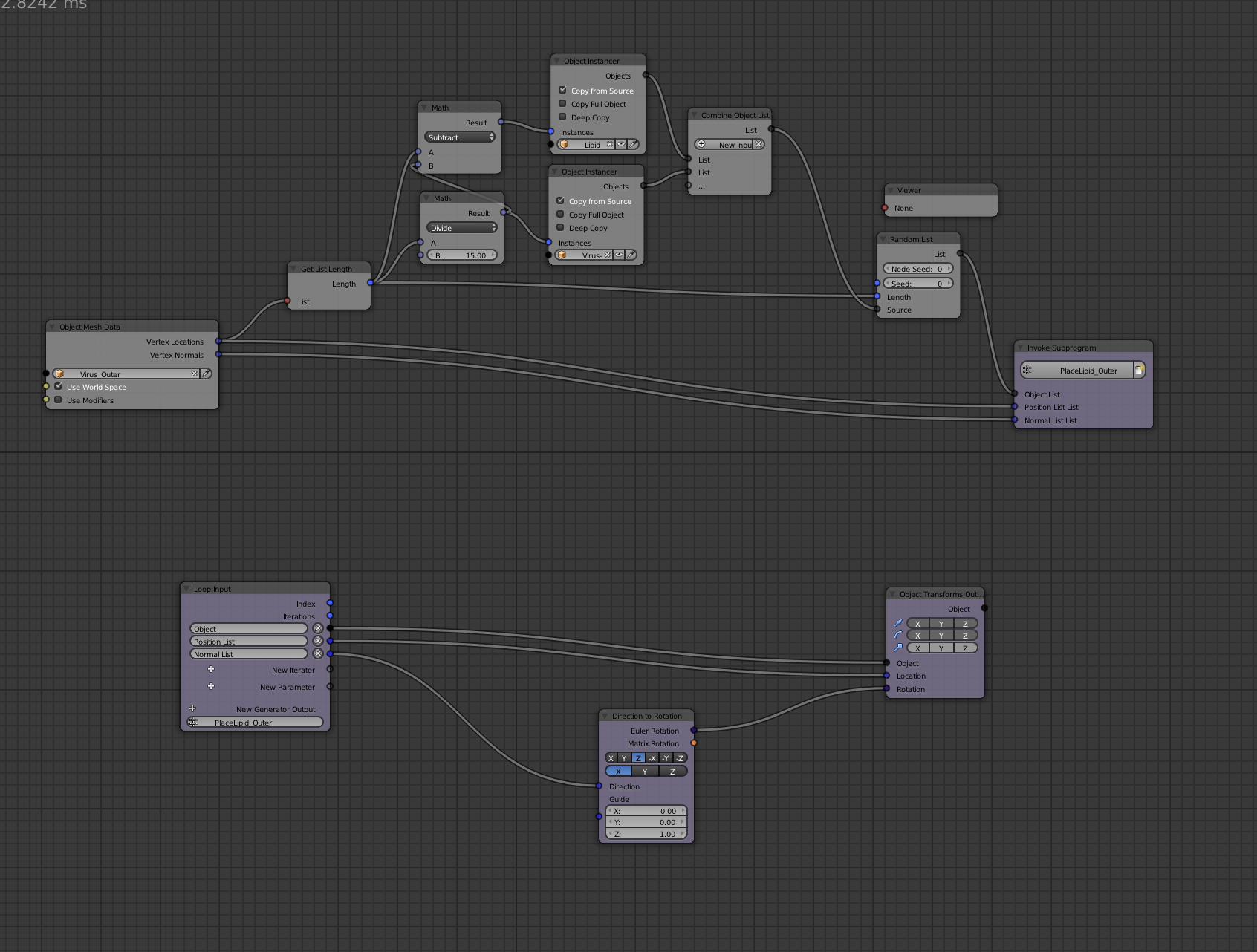
- An animation node network is created to position the lipids and proteins as shown below. The position and the normal for the vertices is used to the lipids/proteins change their orientation when moving.
- To move the lipids/proteins the animation is broken into 4 equivalent parts: the interior of the virus, the exterior of the virus, the top of the cell membrane, and the bottom of the cell membrane. For these 4 parts, the same kind of solution is applied. A source mesh is used to initially place the lipids/proteins. A destination mesh is used to define the location at the end of the animation.
- For the sake of brevity, only the exterior of the virus will be explained in detail. A similar approach is used for the interior, top surface, and bottom surface. The ico sphere for the exterior is created. The mesh is duplicated and edited into a flat surface. This surface is made so the lipids/proteins will follow a simple path from the eco-sphere mesh to the flat mesh.
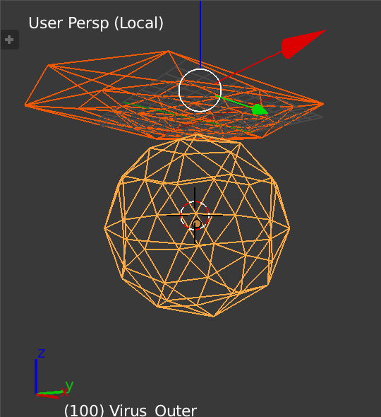
- An animation node network is created to position the lipids and proteins as shown below. The position and the normal for the vertices is used to the lipids/proteins change their orientation when moving.
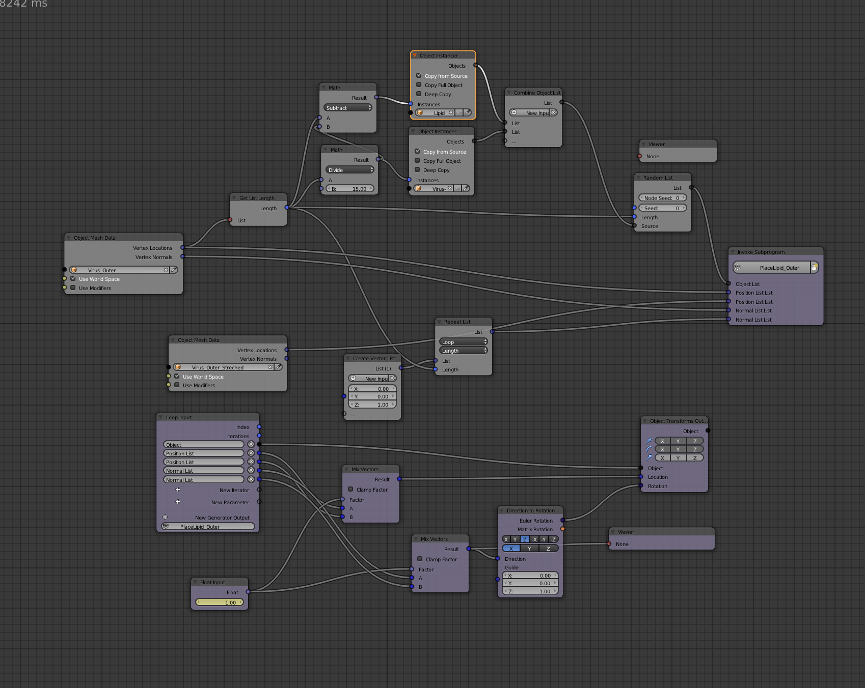
- Similar networks are created for the interior, top, and bottom.
- The meshes for the interior and exterior of the virus are animated moving towards and through the cell membrane. This helps make the merge look smoother.
- The virus contents are animated moving with the interior and exterior meshes and finally into the cell.
Similar networks are created for the interior, top, and bottom.
The meshes for the interior and exterior of the virus are animated moving towards and through the cell membrane. This helps make the merge look smoother.
The virus contents are animated moving with the interior and exterior meshes and finally into the cell.

